Figure 4.
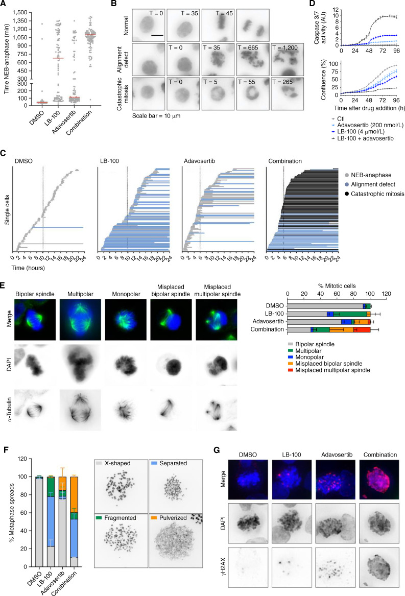
The LB-100 and adavosertib combination leads to aberrant mitoses and cell death. A, Time from nuclear envelope breakdown (NEB) to anaphase for HT-29 cells untreated (DMSO) or treated with LB-100, adavosertib, or the combination. Each dot represents an individual cell followed by live-cell imaging. Red bars represent the average time spent from NEB to anaphase. Two independent experiments are compiled (n =100 cells per condition). B,Representative live-cell microscopy images of HT-29 cells. The examples highlight the two major mitotic phenotypes observed. The scalebar represents 10 μm. C,Representation of the time for mitotic entry and exit of HT-29 cells imaged every 5 minutes for 24 hours, starting immediately after the addition of DMSO, LB-100, adavosertib, or the combination. Each bar represents an individual cell. The colors of the bars indicate normal or aberrant mitoses. The beginning of the bars marks NEB and the end represents either anaphase or end of the experiment. Dashed vertical lines represent the average times for mitotic entry after the addition of the drugs. D,IncuCyte-based assay for caspase-3/7 activity. Cells were treated with DMSO, LB-100, adavosertib, or the combination in the presence of a caspase-3/7 apoptosis assay reagent. Green fluorescence from the apoptosis assay reagent divided by the total confluence was used to estimate apoptosis for 96 hours. E,Representative images (left) and quantification (right) of spindle defects in mitotic cells treated with DMSO, LB-100, adavosertib, or the combination. Cells were treated for 8 hours before fixation. DNA was stained with DAPI (blue) and α-Tubulin was immunostained (green). Quantification is based on 2 independent experiments each analyzing 50 cells per condition. F,Chromosome spreads from HT-29 cells treated with DMSO, LB-100, adavosertib, or the combination. On the left, quantification of chromosome integrity; on the right, are representative images. Drugs were added for 16 hours and then nocodazole was added for an additional hours to block cells in mitosis. Cells were harvested by mitotic shake-off for spreading.G,Representative images show HT-29 cells treated with DMSO, LB-100, adavosertib, or the combination for 24 hours. After fixation, total DNA was stained with DAPI (blue) and γ-H2AX was immunostained (red). Throughout the figure, LB-100 was used at 4 μmol/L and adavosertib at 200 nmol/L.

