Figure S5.
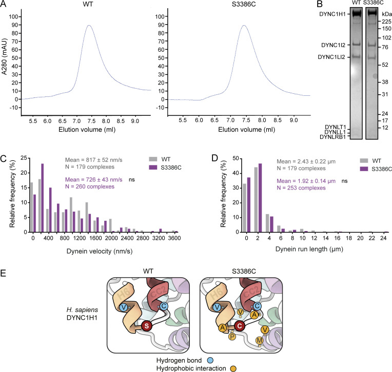
Supplementary data from in vitro and in silico analysis of S3386C human dynein. (A) Size-exclusion chromatography traces for wild-type (WT) and S3386C human dynein complexes, showing very similar profiles and lack of aggregation. (B) Cropped images from a Coomassie Blue–stained SDS-PAGE gel of pooled and concentrated fractions collected from the WT and S3386C mutant dynein peaks shown in panel A. (C and D) Velocity (C) and run length (D) frequency distributions for processive WT and S3386C mutant dynein complexes in the presence of dynactin and BICD2N. Errors are SEM. Evaluation of statistical significance was performed with a Mann-Whitney test. ns, not significant. (E) Zoom-ins of regions of representative examples of WT and S3386C dynein MTBD and stalk MD simulations. Single-letter amino acid codes are shown. Frequent hydrogen bonding interactions of S3386 and C3386 are shown as blue circles and new hydrophobic interactions of C3386 as gold circles; see Tables S1, S2, and S3 for details of interacting residues. Source data are available for this figure: SourceData FS5.

