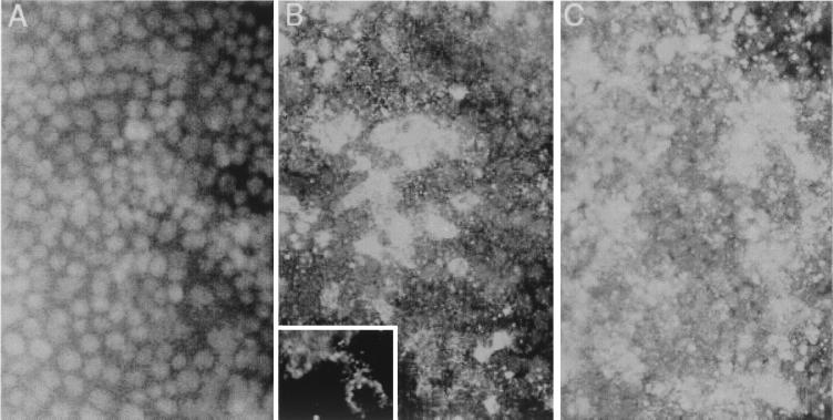
FIG. 2.
Indirect immunofluorescence detection of HAV antigen in infected Caco-2 cells. (A) Normal uninfected Caco-2 monolayer with DAPI nuclear counterstain. (B) Caco-2 cells 8 days following infection with HAV at an MOI of 6.0. The inset shows a high-power view of the cytoplasmic distribution of punctate HAV-specific fluorescence in two cells (no counterstain). (C) Caco-2 cells 12 days following infection with HAV. Viral antigen was visualized with a monoclonal antibody reactive with the virus capsid.

