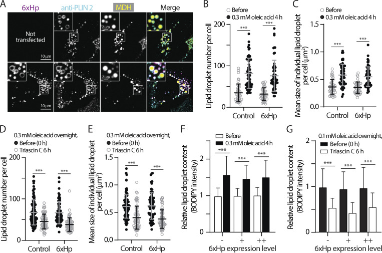
Figure 3.
Minimal perturbation of 6xHp on lipid droplets’ properties. (A) Colocalization of Halo-6xHp and endogenous lipid droplet (LD) protein periplipin 2 (PLIN 2) in an oleic acid (OA)-treated U2OS cell stained with MDH (LD marker) monitored by confocal microscopy. Representative images from a single axial plane (0.3 µm) are shown. (B and C) Number and size of LDs in control HeLa cells or in cells overexpressing Halo-6xHp before and after 0.3 mM OA treatment for 4 h. Mean ± SD are shown (43–59 cells from three or four independent experiments). ***P ≤ 0.001, assessed by one-way ANOVA. (D and E) Number and size of LDs in OA-loaded HeLa cells with or without Halo-6xHp overexpression before and after 10 µM Triacsin C treatment for 6 h. Mean ± SD are shown (62–96 cells from three or four independent experiments). ***P ≤ 0.001, assessed by one-way ANOVA. (F) Relative LD content in HeLa cells overexpressing Halo-6xHp under control conditions or treated with 0.3 mM OA for 4 h. Mean ± SD are shown (98–217 cells from three independent experiments). “−” indicates absence of 6xHp expression. “+” and “++” indicate low and moderate expression of 6xHp, respectively. ***P ≤ 0.001, assessed by one-way ANOVA. (G) Relative LD content in OA-loaded HeLa cells overexpressing Halo-6xHp under control conditions or incubated with 10 µM Triacsin C for 6 h. Mean ± SD is shown (169–499 cells from three independent experiments). “−” indicates absence of 6xHp expression. “+” and “++” indicate low and moderate expression of 6xHp, respectively. ***P ≤ 0.001, assessed by one-way ANOVA.

