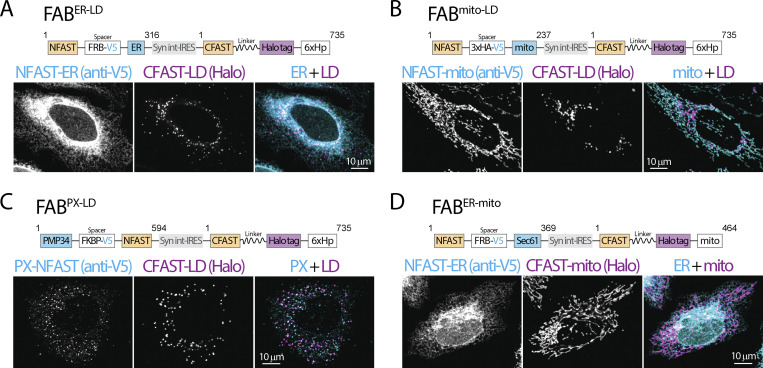
Figure S2.
Diagram and validation of FABCON lentiviruses. (A–D) Diagram (top) and organelle distribution of cognate FABCON halves (bottom) of FABER-LD (A), FABmito-LD (B), FABPX-LD (C), and FABER-mito (D) monitored by confocal microscopy. NFAST fused organelle marker were immunostained with anti-V5 antibody. Maximal intensity projected images from three axial slices (∼1 µm in total thickness) are shown. Numbers of amino acids are indicated. Syn int-IRES, synthetic intron-internal ribosome entry site.

