
Figure 2.
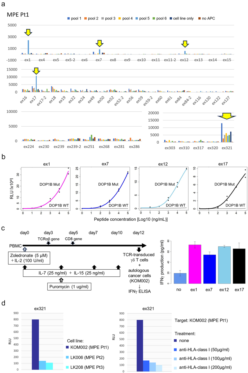
Identification of tumor antigens recognized by CD8+ T cells.
a. Representative data from four separate screenings using 50 TCRs (out of a total 54 TCRs tested) by luciferase reporter assay driven by the NFAT-response element in MPE Pt1. Reactivity of each TCR against six representative pools of peptides (5 peptides/pool) (out of a total 11 pooled). b. Reactivity of the four different TCRs (ex1, ex7, ex12. and ex17) identified in A tested against B-APC pulsed with mutant DOP1B or wild-type peptides at different concentrations. c. Scheme of TCR-transduced γδ T cells co-cultured with the autologous cell line for detection of IFNγ production (left). The results of IFNγ ELISA (right). d. Activation of e×321TCR signaling was assessed against the autologous cell line or other cell lines (LK006 and LK208). The e×321TCR-transduced CD8-J2 cells were also co-cultured with autologous KOM002 with HLA class I-blocking antibody (50, 100 or 200 μg/ml)