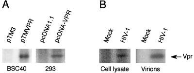
FIG. 2.
Phosphorylation of Vpr in BSC40, HEK293, and HIV-1-infected Jurkat cells. (A). HIV-1 Vpr was expressed from pTM-VPR in BSC40 cells by using the vaccinia virus expression system or pcDNA-VPR in HEK293 cells and labeled with 0.5 mCi of [32P]H3PO4 per ml for 6 h. The cells were harvested, lysed, and immunoprecipitated with anti-Vpr antibodies. Vpr was separated by SDS-PAGE, and the phosphorylated Vpr was detected by exposure of the dried gel to X-ray film. (B) Both HIV-1 p120-infected (7 days postinfection) and noninfected Jurkat cells were labeled with 0.5 mCi of [32P]H3PO4 per ml for 6 h. The cells were harvested, lysed, and immunoprecipitated with anti-Vpr antibodies. The supernatants were passed through 20% sucrose cushions and then immunoprecipitated with anti-Vpr antibodies. The labeled Vpr was detected by exposure of the dried gel to X-ray film and indicated by an arrow.

