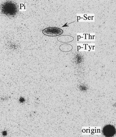
FIG. 3.
PAA of Vpr. The 32P-labeled Vpr was immunoprecipitated from pcDNA-VPR-transfected HEK293 cells, separated by SDS-PAGE, and transferred to an Immobilon-P membrane. The Vpr band was excised and hydrolyzed. The hydrolysate was dried, dissolved in 10 μl of H2O containing 0.5 μg each of phosphoserine (p-Ser), phosphothreonine (p-Thr), and phosphotyrosine (p-Tyr), and then spotted onto a cellulose plate to perform PAA. The three phosphoamino acid locations are shown by ovals; the 32P-phosphoamino acids were revealed by exposure of the plate to a phosphorimager screen.

