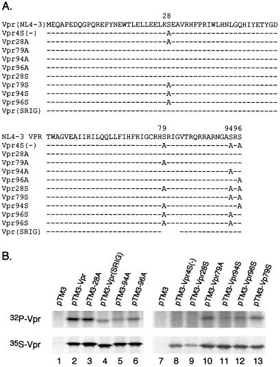
FIG. 4.
Mapping the Vpr phosphorylation sites. (A) Schematic representations of HIV-1 NL4-3 Vpr and various mutations. Serines at positions 28, 79, 94, and 96 were mutated to alanines. Dashes indicate amino acid residues that are the same as in Vpr(NL4-3); Vpr(SRIG) is a SRIG deletion mutant. (B) Wild-type Vpr and relevant mutants were expressed in BSC40 cells using the vaccinia virus expression system. The cells were labeled with 35S or 32P overnight, and labeled Vpr was detected with anti-Vpr antibodies.

