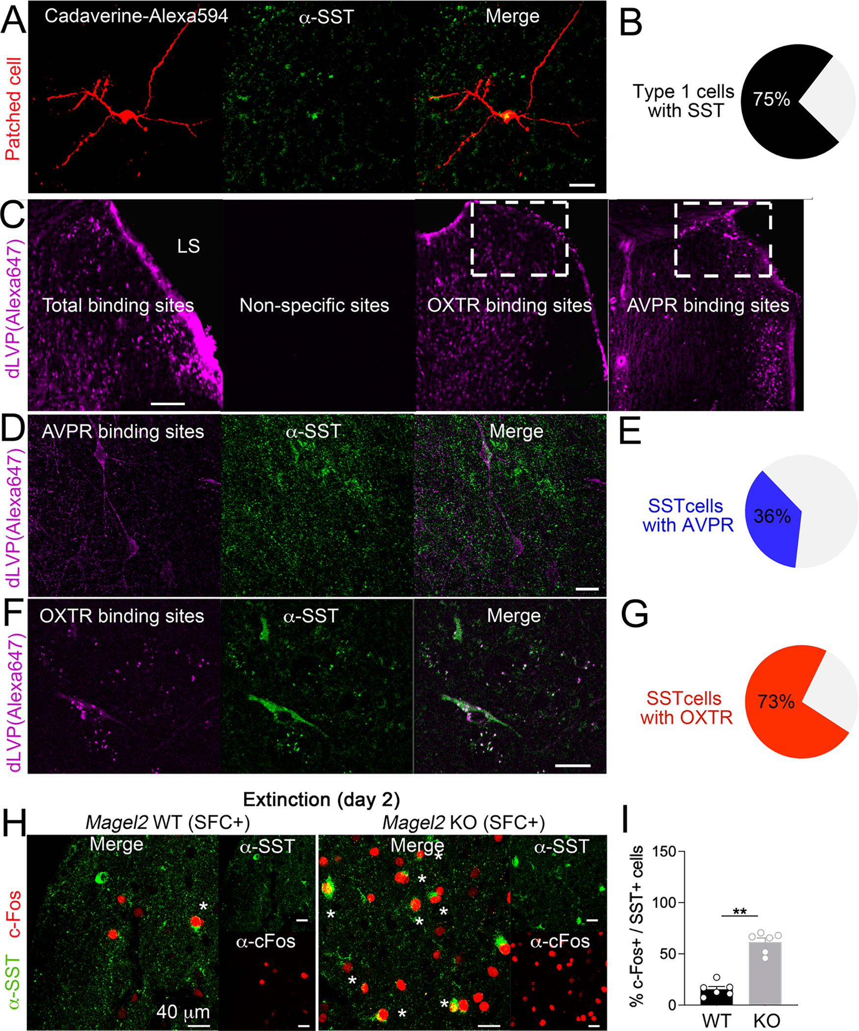
Figure 5. LS neurons modulated by TGOT and AVP are somatostatinergic, and more engaged by social-fear extinction in Magel2KO mice than in WT controls.

(A) Infusion of Cadaverine dye in the patch pipette labeled type-1 cells counterstained with somatostatin (SST) antibodies. Scale=25μm.
(B) Proportion of type-1 cells (N=16) with SST marker. Scale=200μm.
(C) Binding specificity of the fluorescent peptide: 50 μM d[Lys(Alexa-647)8]VP without (total binding) or with 100 μM Manning compound (non-specific). OXTR binding sites marked with 10 μM d[Lys(Alexa-647)8]VP when AVPR were saturated with 5 μM of the competitive ligand Manning compound. AVPR binding sites marked with 50 μM d[Lys(Alexa-647)8]VP when OXTR were saturated with 5 μM of the competitive ligand TGOT.
(D) AVPR binding counter-stained with SST antibodies in dLS. Scale=25μm.
(E) A minority of SST-neurons in dLS (N=203) co-express AVPR binding sites.
(F) OXTR binding counter-stained with SST antibodies in dLS. Scale=25μm.
(G) A majority of SST-neurons in dLS (N=265) co-express OXTR binding sites.
(H) Co-staining with c-Fos and SST antibodies in dLS of Magel2KO mice and WT-controls sacrificed 1h after social-fear extinction.
(I) Proportion of SST-neurons co-labeled with c-Fos in LS. Data are means±SEM in N=6 mice/group. Mann-Whitney test p<0.05.
