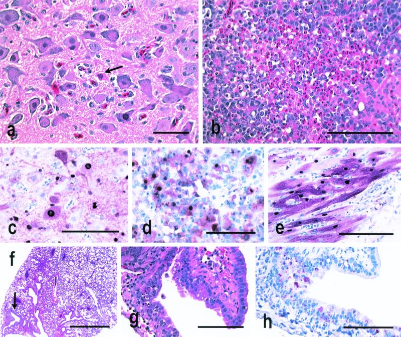
FIG. 3.
Experimental studies in 4-week-old chickens and 7-week-old mice inoculated with A/Env/HK/437-4/99, A/Env/HK/437-6/99, A/Env/HK/437-8/99, and A/Env/HK/437-10/99. Photomicrographs of hematoxylin and eosin-stained tissue sections (a, b, f, and g) and photomicrographs of tissue sections stained immunohistochemically to demonstrate avian influenza virus NP (c to e and h) are shown. (a) Neuronal degeneration and necrosis in the medulla of a chicken euthanized on day 2 after intravenous inoculation with A/Env/HK/437-6/99. Bar, 25 μm. (b) Severe widespread necrosis of pancreatic acinar epithelium from a chicken euthanized on day 2 after intravenous inoculation with A/Env/HK/437-8/99. Bar, 20 μm. (c) Intranuclear and intracytoplasmic avian influenza virus antigen in neurons and glial cells from the chicken in panel a. Bar, 50 μm. (d) Intense staining of pancreatic acinar epithelium and debris for avian influenza virus antigen from the chicken in panel b. Bar, 50 μm. (e) Intranuclear and intracytoplasmic avian influenza virus antigen in cardiac myocytes of a chicken that died on day 2 after intravenous inoculation with A/Env/HK/437-10/99. Bar, 50 μm. (f) Single focal area of bronchitis in a normal lung from a mouse euthanized on day 4 after intranasal inoculation with A/Env/HK/437-4/99. Bar, 500 μm. (g) Higher magnification of panel f, showing focal necrosis of respiratory epithelium from a bronchus. Bar, 50 μm. (h) Avian influenza virus antigen in bronchial respiratory epithelium of the mouse in panel f. Bar, 50 μm.

