
FIGURE 3.
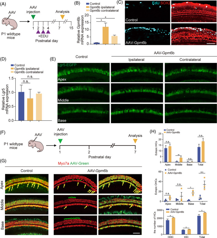
Gpm6b overexpression promoted hair cell regeneration in vivo. (A) Experimental design. (B) mRNA expression level of Gpm6b in P15 cochleae after the injection of AAV‐Gpm6b. (C) EdU immunostaining after Gpm6b overexpression in cochlear. EdU (cyan) marks proliferating cells. Sox2 (red) marks the SCs. Scale bar: 50 μm. (D) mRNA expression level of Lgr5 in P15 cochleae after the injection of AAV‐Gpm6b. (E) Immunofluorescence images of the AAV‐transduced cochleae of Lgr5‐EGFP mice. Lgr5‐EGFP (green) marks the Lgr5+ cells. (F) Experimental design. Tamoxifen was injected intraperitoneally into P1 wildtype mice. The AAVs were injected through the round window membrane, and the cochleae were collected at P7. (G) Immunofluorescence images of cochlear epithelia infected by control and AAV‐Gpm6b, respectively. Ectopic HCs are marked by yellow arrows (IHC area) and green arrows (OHC area). Scale bar: 50 μm. (H) The number of ectopic IHCs, ectopic OHCs, and total HCs in (B). AAV dose: 2.8 × 10¹⁰ GCs per cochlea. Data are displayed as the mean with SEM. The value of p was calculated by Student's t‐test. *p < 0.05; **p < 0.01; ***p < 0.001; n.s. refers to no significance.