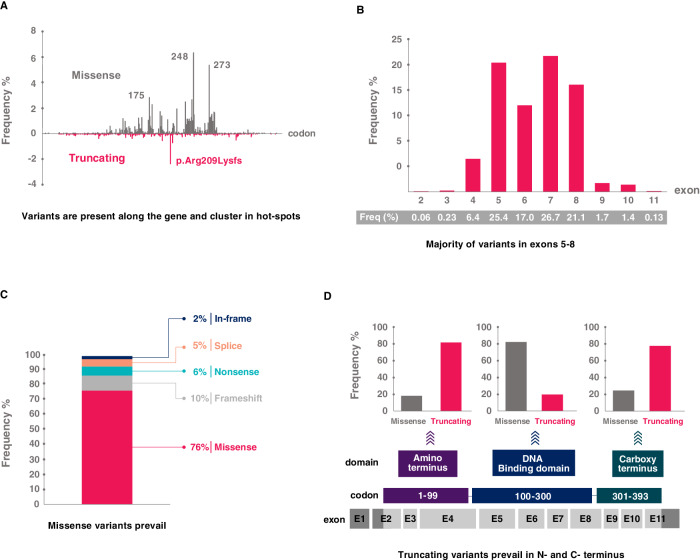
Fig. 4. Spectrum of TP53 defects detected in CLL.
TP53 variant profile based on data collected for CLL patients in the UMD database; common polymorphisms have been omitted [82]. A Codon distribution with hot-spot variants depicted. Variants in codons 175, 248, and 273 are general hot spots, while the truncating frameshift variant in codon 209 is CLL-specific. B Exon distribution showing the prevalence of variants in exons 5–8. C Proportion of variant types out of all variants. D Proportion of variant types in individual domains. In the DNA-binding domain, missense variants prevail; conversely, truncating variants are predominant in the carboxy and amino termini.

