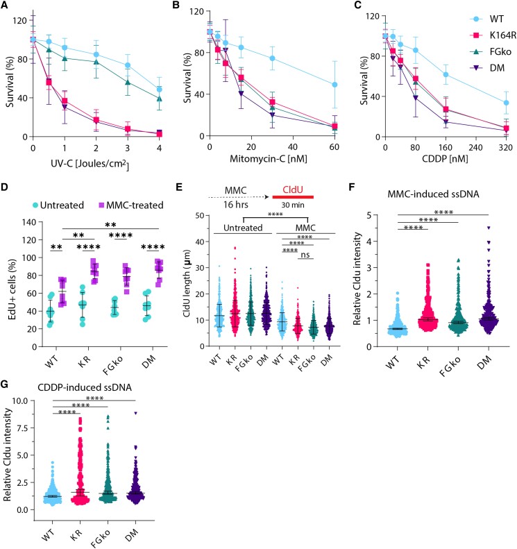
Fig. 3.
PCNA-Ub is a central player in crosslink repair. A–C) Survival of WT, KR, FGko, and DM Trp53kd MEFs obtained by colony formation assay. Cells were seeded at increasing densities in 10-cm dishes, treated with increasing doses of A) UV-C, B) CDDP, and C) MMC and fixed, stained, and counted on day 7 after seeding. Colonies were counted using a colony counter from three independent experiments. For each dose, graph displays mean ± SD. D) Percentage of EdU-positive cells in untreated or MMC-treated MEFs. Each dot indicates a sample, and bars represent mean ± SD. Cells were left untreated or treated with 1.5 µM MMC for 16 hours followed by EdU labeling for 20 minutes before being harvested and fixed in 70% ethanol. In addition, the DNA stain DAPI was used as a cell cycle marker to distinguish G1 from G2/M cells. E) Length of CldU-labeled DNA fibers. Cells were left untreated or treated with 1.5 µM MMC for 16 hours followed by CldU labeling for 30 minutes before being harvested. At least 400 fibers were measured. Bars represent mean ± SD. P-values were calculated using one-way ANOVA. **P < 0.01, ****P < 0.0001. F), G) Quantification of CldU-based ssDNA based on protocol from Koundrioukoff et al. (47). Cells were grown in the presence of CldU for 24 hours before treatment or not with F) 1.5 µM MMC or G) 8 µM CDDP for 16 hours prior to fixation. CldU was immuno-detected without DNA denaturation, which only permits visualization of single-stranded regions. Nuclei were counterstained with DAPI. Bars represent median ± 95% CI.

