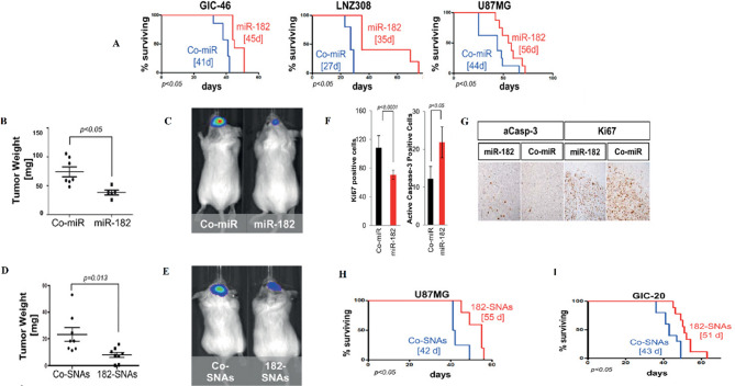
Fig. 22.
(A) Survival analysis indicated that miR-182 expression increased the survival of animals (rthotopic xenograft with glioma cells and engineered GICs that stably expressed miR-182). (B and C) Tumor burden analysis via weight and bioluminescence imaging. (D) Weight of tumors derived from U87MG xenografts extracted from SCID mice 21 days after intravenous treatment with Co-SNAs or 182-SNAs. (E) Bioluminescence imaging of xenograft tumors derived from GIC-20 (12 day) after intravenous treatment with Co-SNAs or 182-SNAs. (F) Estimation levels of Ki67 and caspase-3 in xenograft samples. (G) Ki67 and caspase-3 IHC in coronal brain sections of GIC-derived xenografts expressing Co-miR or miR-182. (H and I) Kaplan-Meyer survival estimator curves of SCID mice xenografted with glioma tumors (U87MG and GIC-20) and intravenously treated with Co-SNAs or 182-SNAs.
Permission was received from ref [160]

