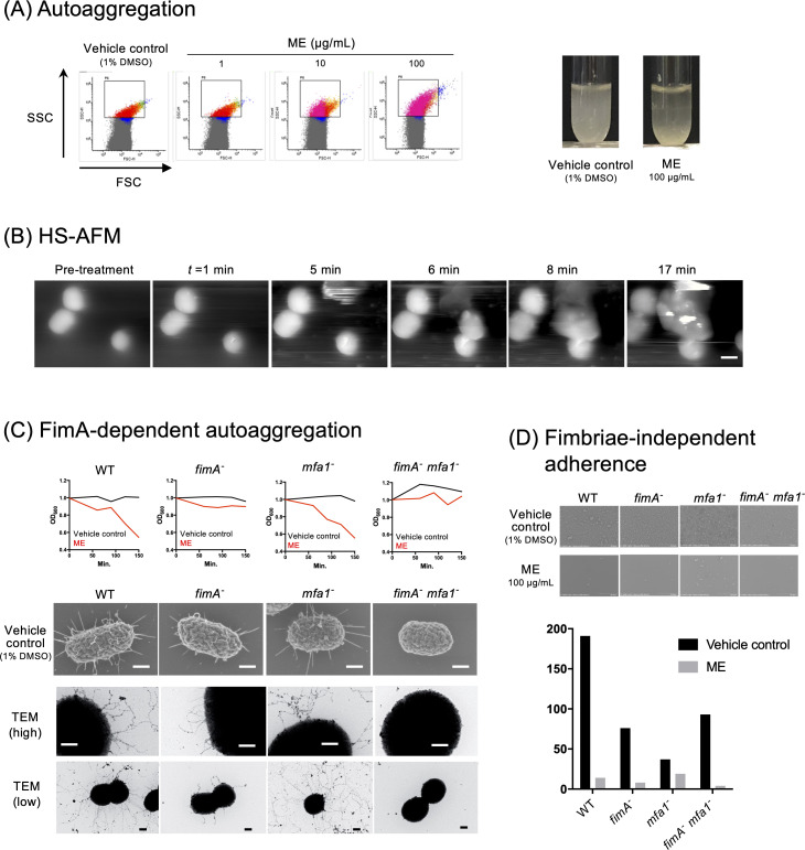
Fig 2.
Aggregation and adherence of P. gingivalis treated with ME. (A) In vitro autoaggregation assay. In flow cytometry analysis (left), P. gingivalis cells were treated with vehicle control (1% DMSO) or ME at different concentrations (1, 10, and 100 µg/mL). The x- and y-axes showed the FSC and the SSC, respectively. Shown (right) are the difference in turbidity of the tubes 2 h after treatment with vehicle control (1% DMSO) and ME (100 µg/mL) in in vitro tube assay. (B) Real-time HS-AFM imaging of aggregation of P. gingivalis cells after ME treatment (1.0 mg/mL). Bar: 300 nm. See also Video S2. (C) FimA-dependent autoaggregation. In vitro aggregation assays were performed using P. gingivalis WT, FimA mutant (fimA−), Mfa1 mutant (mfa1−), and FimA and Mfa1 double mutant (fimA− mfa1−) strains in the presence or absence of ME. The top panels showed the change in turbidity (OD600) for 150 min. The morphology of each P. gingivalis cell (FE-SEM) treated without or with ME at a concentration of 1 mg/mL is also shown in the middle or bottom panel, respectively. Bars: 200 nm. (D) Fimbriae-independent adherence. ME dramatically decreased the adherence of cells not only of wild type but also of a series of fimbrial mutant strains.

