
Fig 2.
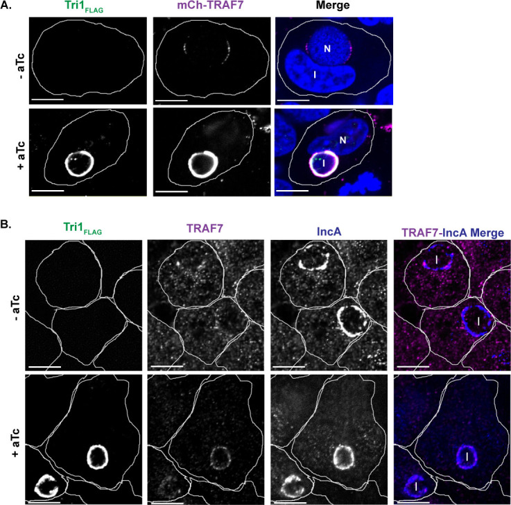
TRAF7 is recruited to the inclusion. (A) Confocal immunofluorescence microscopy of HeLa cells transfected with mCh-TRAF7 for 24 h and then infected with L2+pTri1FLAG for 24 h with or without aTc induction. Cells were fixed and stained with α-FLAG to visualize Tri1FLAG. Merged images show mCh-TRAF7 (pseudo-colored magenta), Tri1FLAG (pseudo-colored green), and DAPI (4′,6-diamidino-2-phenylindole) (blue) staining. (B) Confocal immunofluorescence microscopy of HeLa cells infected with L2+pTri1FLAG for 24 h with or without aTc induction. Cells were fixed and stained with antibodies to FLAG (to detect Tri1), TRAF7 (pseudo-colored magenta in merge), and IncA (blue in merge, to delineate the inclusion membrane). The merge panel only includes TRAF7 and IncA. The small amount of transfected mCh-TRAF7 present at the inclusion in the absence of inducer likely represents recruitment by chromosomally encoded Tri1. Shown are single z-slices. I, inclusion. N, nucleus. Scale bar = 10 µm. Cell outlines are included for clarity.