Figure 6.
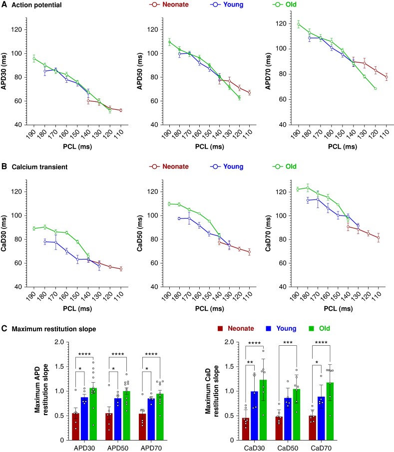
Cardiac electrical restitution properties in response to extrasystolic stimulation. A) Action potential duration restitution curves at APD30, APD50, and APD70 in neonatal (n = 5–6), younger adult (n = 5), and older adult hearts (n = 6–11). B) Calcium transient duration restitution curves at CaD30, CaD50, and CaD70 in neonatal (n = 5–6), younger adult (n = 5), and older adult hearts (n = 6). C) Maximum restitution slopes; individual replicates shown. Extrasystolic stimulation was applied to the ventricle (S1–S2) at the apex. Note the different PCL ranges that were achievable in neonatal vs. adult hearts. Values reported as mean ± SEM. Statistical comparisons between slope measurements, as determined by one-way ANOVA with multiple comparisons; *P < 0.05, **P < 0.01. APD, action potential duration; CaD, calcium transient duration; PCL, pacing cycle length.

