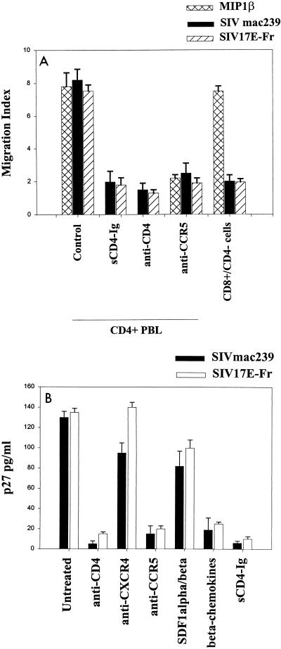
FIG. 1.
Exposure of PHA–IL-2-activated pigtail macaque PBMCs to SIVmac239 or SIV 17E-Fr causes chemotaxis (A) and infection (B). Chemotaxis to SIV or MIP1-β was inhibited by pretreatment of cells with 10 μg of anti-CD4 MAb (Leu3A) or anti-CCR5 MAb (2D7) per ml for 30 min on ice. Alternatively, purified virus was pretreated with sCD4-Ig (10 μg/ml) for 30 min on ice before use in the chemotaxis assay, as described in Materials and Methods. Macaque CD8+ cells isolated by negative depletion with anti-CD4 magnetic beads (>85% CD8+, <2% CD4+ cells) did not migrate in response to SIVmac239 or SIV 17E-Fr. CD4+ and CD8+ lymphocytes expressed comparable levels of CXCR4 and CCR5 and had similar migratory dose-response curves to SDF1-α and MIP1-β. Infection with 103 50% tissue culture infective doses of SIVmac239 or SIV 17E-Fr was inhibited by pretreatment of cells with 10 μg of MAbs to CD4 or CCR5 per ml or 200 ng of the beta chemokines (RANTES, MIP1-α and MIP1-β) per ml for 30 min on ice but not with anti-CXCR4 MAb (12G5) or 200 ng of the alpha chemokines (SDF1-α and SDF1-β) per ml. Treatment of purified virus with sCD4-Ig for 30 min on ice prior to infection of cells was also inhibitory. Supernatants were assayed for p27 on day 7. Results represent the mean ± standard error of the mean of three experiments. Similar trends were observed on days 10 and 14, although absolute values of p27 were higher.

