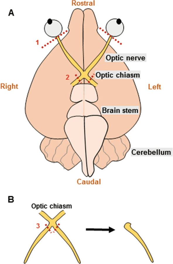
Fig. 1.

(a) Schematic showing the optic nerves attached at the optic chiasm located at the base of the brain. During dissection to isolate the nerves, the right and left optic nerves are cut right behind the retina (#1), following which the brain is lifted out of the skull and the optic chiasm is removed from the brain (#2). (b) Optic nerves are transferred to the recording chamber and cut twice under a dissecting microscope (#3) to separate the right and left optic nerves for recordings
