Fig. 3.
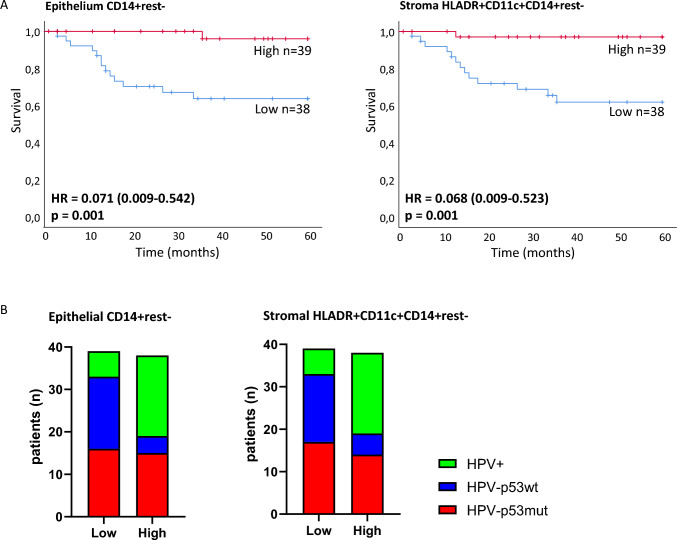
Pro-inflammatory myeloid cell composition predicts excellent survival in VSCC. A Kaplan–Meier survival curves for VSCC (n = 77) with high or low myeloid cell infiltration, including hazard ratio (HR, between brackets the 95% confidence interval of the hazard ratio is provided), and p-value. Total VSCC cohort split by median into high versus low counts. Left survival curve for separation by CD14+rest− monocyte infiltration, low infiltration (blue): HPV−p53mut n = 16, HPV−p53wt n = 17, HPV+ n = 6, high infiltration (red): HPV−p53mut n = 15, HPV−p53wt n = 4, HPV+ n = 19. Right survival curve for separation by stromal HLADR+CD11c+CD14+rest− dendritic cell infiltration, low infiltration (blue): HPV−p53mut n = 17, HPV−p53wt n = 16, HPV+ n = 6, high infiltration (red): HPV−p53mut n = 14, HPV−p53wt n = 5, HPV+ n = 19. B Distribution of VSCC molecular subtypes (n = 77 total) across low versus high (split by median) epithelial CD14+rest− monocyte infiltration (left graph), and stromal HLADR+CD11c+CD14+rest.− dendritic cell infiltration (right graph)

