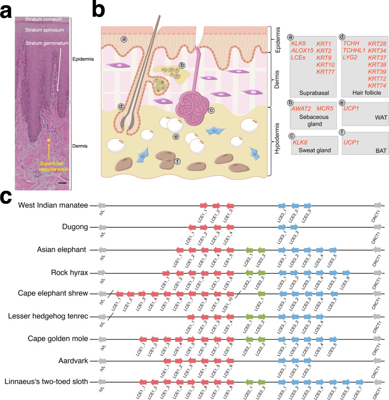
Fig. 3. Anatomy and gene changes of the sirenian integumentary system.
a Representative histology of hematoxylin and eosin (H&E)-stained dorsal dugong skin. The scale bar represents 50 μm. b Schematic drawing of H&E-stained human skin (as a model of mammals) and associated genes with changes in sirenians. Left, overview of major skin anatomical structures: epidermis, dermis, hypodermis, and ectodermal appendages (hair follicles, sebaceous glands, and sweat glands). Right, genes and their site of expression. Inactivated genes are in red (see main text). WAT denotes white adipose tissue cell; BAT brown adipose tissue cell. c Schematic representation of late cornified envelope (LCE) gene clusters in afrotherian genomes. Arrows indicate genes and their direction of transcription; slashes indicate separate scaffolds; and different clusters are indicated by colored boxes.

