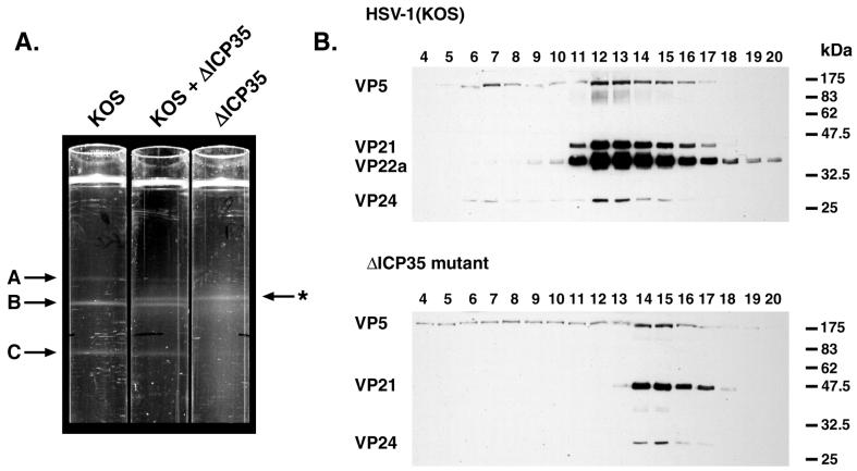
FIG. 4.
Altered sucrose gradient sedimentation of mutant capsids. (A) The positions of light-scattering bands corresponding to wild-type A, B, and C capsids and ΔICP35 mutant B capsids (marked with an asterisk) are indicated by arrows adjacent to three 20 to 50% (wt/vol) sucrose gradients centrifuged in parallel. The gradients contain lysates from HSV-1 (KOS)- and ΔICP35-infected cells, as indicated. (B) Fractionated sucrose gradients of HSV-1 and ΔICP35 mutant virus-infected cells were analyzed by Western blotting with antibodies against VP5, VP21/VP22a, and VP24, as indicated. Fractions were collected beginning from the bottom of the gradient (fraction 1) to the top (fraction 20). Fractions 4 through 20 are shown. The positions of molecular size standards electrophoresed in parallel are indicated on the right (in kilodaltons). Photographs were prepared for presentation using a Kodak DCS400 digital camera and Adobe PhotoShop 5.0.

