. 2024 May 14;20(4):459–461. doi: 10.3988/jcn.2023.0481

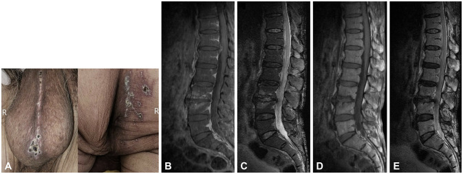
Fig. 1. Dermatologic examination and lumbosacral spine magnetic resonance imaging (MRI). Unilateral vesicular rashes in its healing stage distributed over the S2 and S3 dermatomes on the right buttocks and perineal raphe (A). Contrast-enhanced sagittal fat-suppressed T1-weighted image (B), sagittal fat-suppressed T2-weighted image (C), sagittal T1-weighted image (D), and sagittal T2-weighted image of the lumbosacral spine in MRI (E). There was no definite abnormal enhancement along the cauda equina, except for questionable signal alterations around the whole spinal cord and spinal canal, which required a differential diagnosis between an artifact and true myelopathy.

Fig. 1