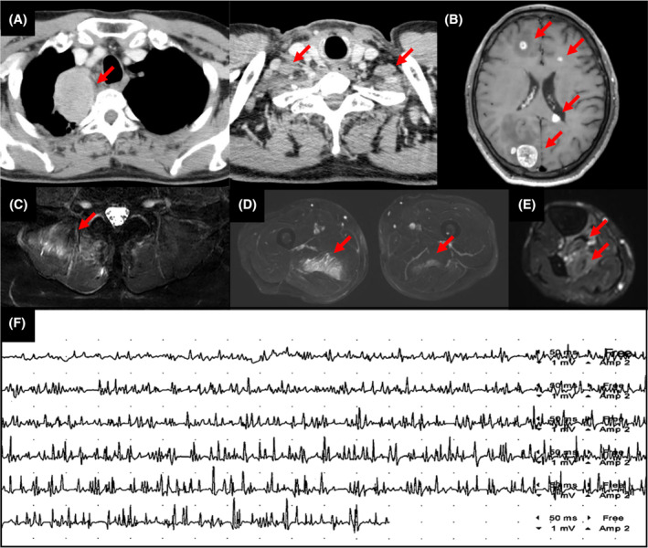
FIGURE 1.

Diagnostic images of a 59‐year‐old male patient with lung adenocarcinoma. (A) Contrast‐enhanced computed tomography image shows lung cancer in the right upper lobe and bilateral supraclavicular lymphadenopathy. (B) Magnetic resonance imaging reveals multiple brain metastases. Axial STIR images shows hyperintense signals in the (C) right erector spinae muscles, (D) bilateral adductor magnus muscle, (E) right posterior tibial muscle, flexor digitorum longus muscle, and flexor hallucis longus (arrows). (F) Electromyography shows early recruitment and polyphasic, short‐duration, low‐amplitude motor unit action potentials.
