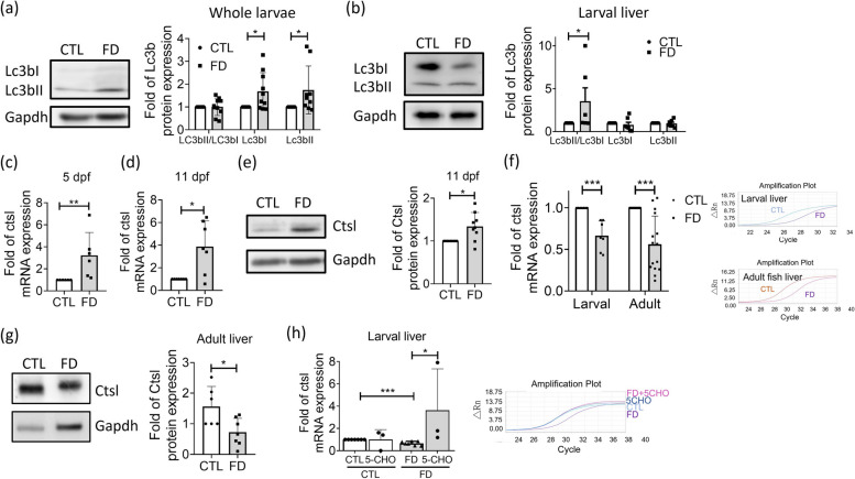
Fig. 10.
Up-/dysregulation of autophagy and down-regulation of cathepsin L expression also occurred in the liver of FD fish in vivo. a-b Cell lysates prepared from the whole larvae (a) and isolated liver from larvae of 11 dpf (b) were Western blotted for Lc3b and quantified with densitometry. Elevated Lc3bII/Lc3bI ratio was most apparent in FD larval liver, signifying an enhanced autophagosome formation, interrupted autophagosome-lysosome fusion, or lysosomal deregulation. Data were collected from at least 7 independent experiments. (c-d) Real-time PCR performed with the sample prepared from whole larvae of 5 dpf (c) and 11 dpf (d) revealed increased cathepsin L expression. Data were collected from at least 6 independent experiments. e Western blotting was conducted with the extract prepared from the whole larvae of 11 dpf for cathepsin L protein level, which revealed an increased expression for FD group. Data were collected from 9 independent experiments. f-g The liver-specific decrease in the expression of cathepsin L was observed both in the larvae of 11 dpf and adult fish of FD groups for both mRNA (f) and protein (g) levels. Data were collected from at least 6 independent experiments. h Larvae were induced for FD and supplementing with 1 mM 5-CHO-THF from 7 to 11 dpf before collected and examined for cathepsin L expression. Folate supplementation significantly increased the cathepsin L mRNA in FD larval liver. Presented are the averaged results of at least three independent trials with each of the sample prepared from 10-20 larvae. CTL, control (Tg-GGH/LR larvae without FD); FD, folate deficiency; 5-CHO, 5-formyl-THF. Statistical data are shown in mean ± SEM. * p<0.05, **, p <0.01; ***, p<0.001

