Fig. 9.
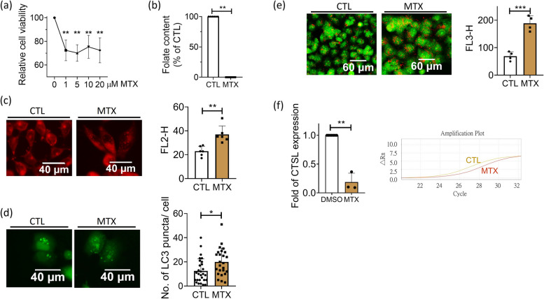
The folate deficiency caused by methotrexate altered autophagic activity and decreased cathepsin L expression in Huh7 cells. a Huh7 cells were examined for viability after cultivating in regular DMEM medium containing methotrexate (MTX) of indicated concentrations. An approximately 30% decrease in cell viability was observed in the presence of MTX at 1 µM and beyond. Data were collected from 5 independent experiments. b An almost complete depletion of intracellular folate content was reached after cells were cultivated in 5 µM MTX for 2 days. Data were collected from 6 independent experiments. c-e Cells grown in 5 µM MTX for 2 days were stained with Nile red for lipid deposition (c), transfected with plasmids encoding LC3-eGFP for characterizing autophagic activity (d) and subjected to Acridine orange staining for acidic vesicles (e). Increased lipid accumulation, autophagy and lysosomes/autolysosomes were apparent in MTX-treated cells. Data were collected from at least 3 independent experiments. f Cells were harvested 2 days after exposing to 5 µM MTX and examined for cathepsin L mRNA. A significant decrease in cathepsin L expression was found for MTX-treated cells. Presented are the averaged results of at least three independent trials. MTX, methotrexate. Statistical data are shown in mean ± SEM. * p<0.05, **, p <0.01; ***, p<0.001

