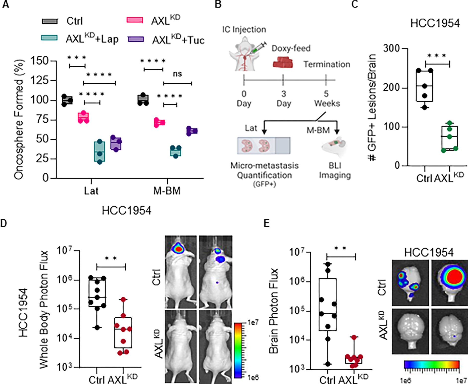
Figure 3. AXL depletion attenuates latent and metachronous brain metastasis.

A, Relative quantification of oncospheres formed by HCC1954 Lat and M-BM cells upon AXL knockdown (AXLKD) and Lapatinib (1μM) or Tucatinib (3μM) treatment for seven days. Data are presented as mean ± SEM. One-way ANOVA was used, followed by the Dunnett test. *, P < 0.05, ***, P < 0.001, ****, P < 0.0001, ns: not significant. B, Experimental scheme to assess the impact of AXL depletion on metastatic latency and relapse. C, Quantification of GFP+ brain metastatic lesions in mice bearing HCC1954 AXLKD Lat cells (n = 5 per group). Mice were on doxycycline feed 3 days post injection until study termination. Data are presented as mean ± SEM. Mann–Whitney U test, ***, P < 0.001. D-E, Whole body and ex-vivo brain metastasis incidence as measured by BLI signal from athymic nude mice bearing HCC1954 AXLKD M-BM cells. (Ctrl group n = 9 and AXLKD group n = 8). Mice were on doxycycline feed 3 days post injection until study termination. Data are presented as mean ± SEM. Mann–Whitney U test, **, P < 0.01. Two representative BLI images of whole-body mice and ex-vivo brains of their respective conditions. (B, Created with BioRender.com).
