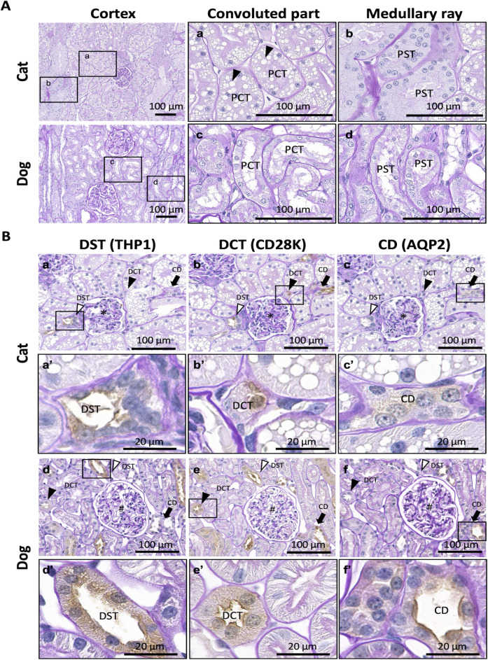
Fig 1. Histological features of renal cortices in adult cats and dogs.
(A) Proximal tubules. Proximal convoluted tubules (PCTs) and proximal straight tubules (PSTs) are observed in the convoluted and medullary regions, respectively. Panels a–d magnified the squared areas in the left panels. Arrowheads denote abundant lipid droplets (LDs) in the cytoplasm of cat PCT epithelial cells. Periodic acid Schiff-hematoxylin (PAS-H) staining. (B) Distal tubules (DTs) and collecting ducts (CDs). The squared areas in panels a–f are magnified in panels a’–f’, serial sections for each animal. Asterisks and sharps indicate glomeruli (panels a–c and d–f, respectively). In both species, Tamm–Horsfall protein 1 (THP1) is positive in the distal straight tubules (DSTs; white arrowheads) but not in the distal convoluted tubules (DCTs; black arrowheads) and CDs (black arrows) (panels a, a’, d, and d’). Calbindin-D28K (CD28K) is positive for DSTs, DCTs, and CDs (panels b, b’, e, and e’). Aquaporin 2 (AQP2) is positive in CDs (black arrows) but not in DSTs and DCTs (panels c, c’, f, and f’). Immunohistochemistry was performed using PAS-H staining. Resolution of anatomical or histological images: 300 × 300 dpi.

