
Extended Data Fig. 8. Nitrate reduces zinc levels in nodules and zinc promotes FUN condensates in the nucleus.
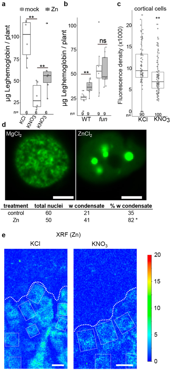
a-b The leghemoglobin corresponding to ARA in Fig. 4f,g. ns: not significant. c Nitrate exposure triggers a reduction in cellular zinc levels within nodules as indicated by the Zinpyr-1 fluorescent dye at 24 h post treatment. The average intensity of the cortical cell zone of nodules. The Box plots show Min, Q1, Median, Q3, Max and individual values (dots) in a-c. Significant differences are determined by ANOVA and Tukey post-hoc testing (**: p value < 0.01; *: p value < 0.05). Biological independent samples n value shown on each box plots and bar plots. d Co-infiltration with ZnCl2 increases the frequency of FUN nuclear condensates in N. benthamiana leaves. Leaves were infiltrated with Agrobacterium carrying a binary vector to express pro35S:FUN-GFP and subsequently infiltrated with 500 µM MgCl2 or ZnCl2 two days before confocal observation. Chi-squared testing (*: p value < 0.05). Scale bar 5 µm. e XRF regions used for quantification. Regions analysed for quantification of zinc from Fig. 4f are boxed. The dash lines indicate the boundaries between nodule cortex (above line) and infected region (below line). Scale bar 20 µm.