
Fig. 4. TET1 protects hPGCLCs from differentiation into extraembryonic cells.
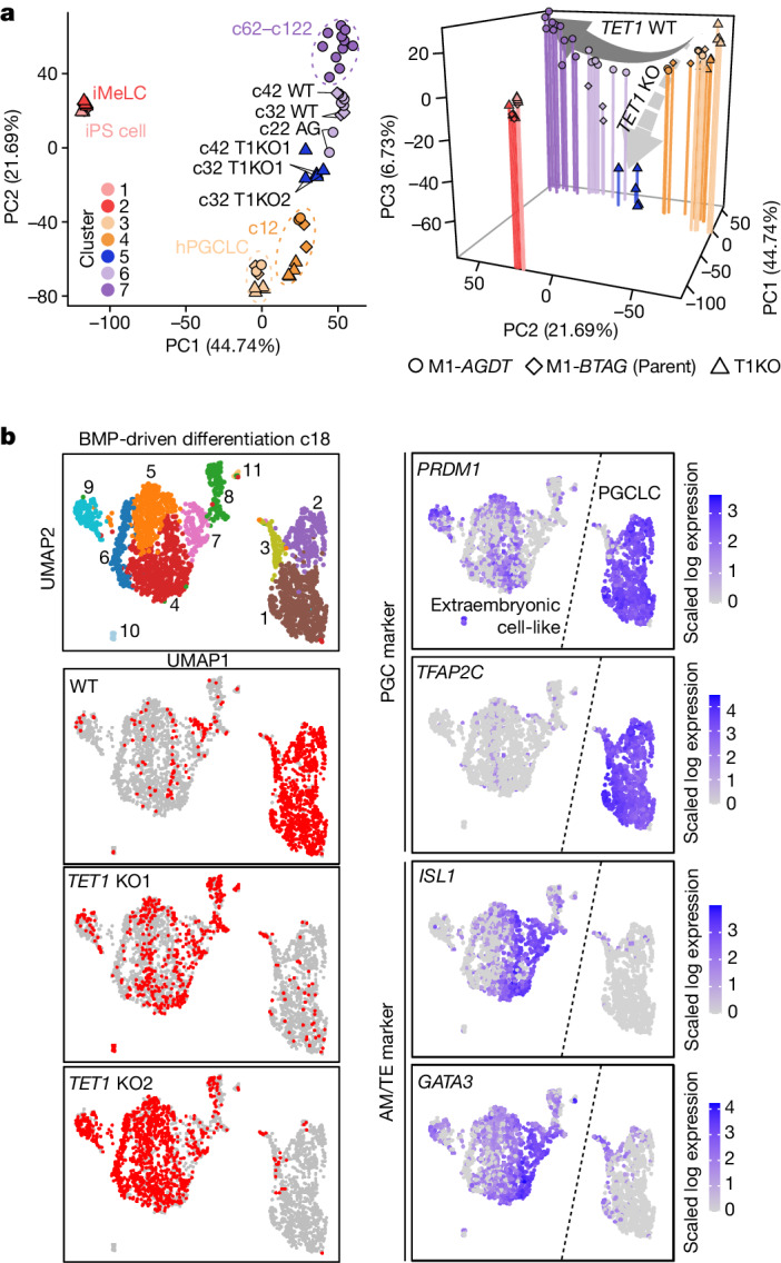
a, PCA (left: PC1 and PC2; right: PC1, PC2 and PC3) of the transcriptomes of BMP-driven wild-type (WT) cell and TET1 KO (T1KO) hPGCLC differentiation (see Supplementary Table 2 for full sample information and Extended Data Fig. 10h for cluster information). Colour coding is as indicated. b, UMAP and Louvain clustering of scRNA-seq data of wild-type and TET1 KO hPGCLC culture at c18 (left column, top), with the annotation of the genotype (left column) or with the expression levels of the indicated genes (right column). Colour code is as indicated.