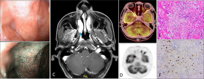
Figure 1.
Fibroscopy, radiologic, and histologic features of IgG4-RD patient. (A–C) Fibroscopy and MRI suggest bulging of the right nasopharynx, disappearance of the crypt, and shallow pharyngeal opening of the eustachian tube. Note the involvement of the fatty gap of the right petrostaphylinus. (D) PET-CT showed active FDG metabolism at nasopharynx. (E, F) Hematoxylin-eosin staining demonstrating lymphocytes, plasma cells, and a few neutrophils, eosinophils infiltrate, accompanied by fibrous tissue hyperplasia. Immunohistochemical staining showed IgG4-positive cells, greater than 10/HPF. These images are at a 20X original magnification.

