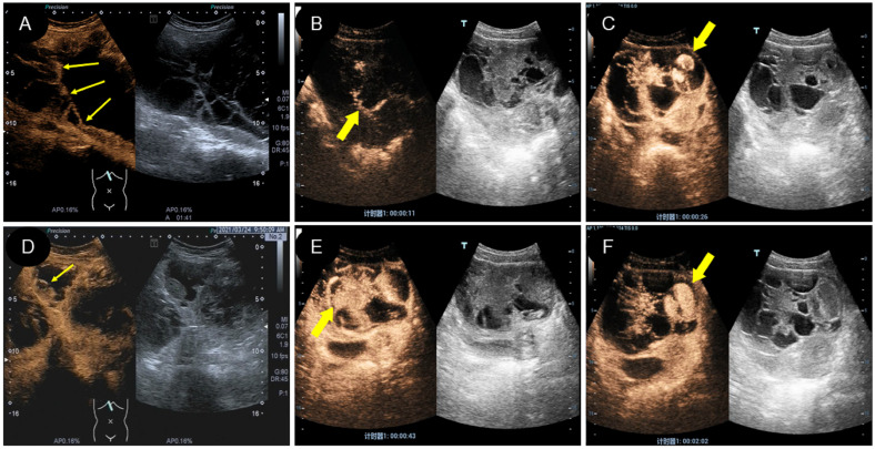
Figure 5.
Characteristics of CEUS different types of yolk sac tumor. (A, D). A mixed yolk sac tumor showed rapid contrast enhancement and no contrast perfusion in the sac (thin arrow); (B, E). A solid yolk sac tumor showed rapid enhancement of solid and cystic area separation, with large internal vessels in the early stage (thick arrow) and uniform high enhancement after peak (thick arrow); (C, F). In this case of a solid yolk sac tumor, the contrast agent flooded into and accumulated into the sac wall (thick arrow) after a burst, leaving a large number of contrast agent remained in the cystic cavity (thick arrow) after the resolution of the solid part.

