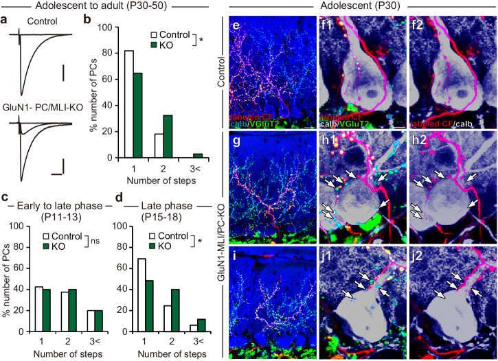
Fig. 6. NMDA receptors in MLIs are involved in CF synapse elimination.
a, b Impaired CF synapse elimination in GluN1-MLI/PC-KO mice aged P30 to P50. Sample CF-EPSC traces (a) and frequency distribution histograms in terms of the number of discrete CF-EPSC steps (b) in control (white columns) and GluN1-MLI/PC-KO (green columns) mice. Holding potential, − 10 mV. Scale bars, 10 ms and 0.5 nA. *P < 0.05, Mann-Whitney U test. c, d Developmental changes in CF innervation. Frequency distribution histograms of PCs in terms of the number of discrete CF-EPSC steps during P11−P13 (c), and P15−P18 (d) in control (white columns) and GluN1-MLI/PC-KO (green columns) mice. *P < 0.05, Mann-Whitney U test. e, f Triple immunofluorescence labeling for calbindin (blue or gray), anterogradely labeled (DA488-positive) CFs (red), and VGluT2 (green) in a control mouse at P30. Scale bar: 20 μm for (e), 5 μm for (f). g, h Triple immunofluorescence labeling similar to (e) and (f) but for data from a GluN1-MLI/PC-KO mouse. i, j Triple immunofluorescence labeling similar to (g) and (h) but for data from another GluN1-MLI/PC-KO mouse. Arrows in (h) and (j) indicate anterogradely unlabeled (DA488-negative) VGluT2-positive CF terminals on PCs.

