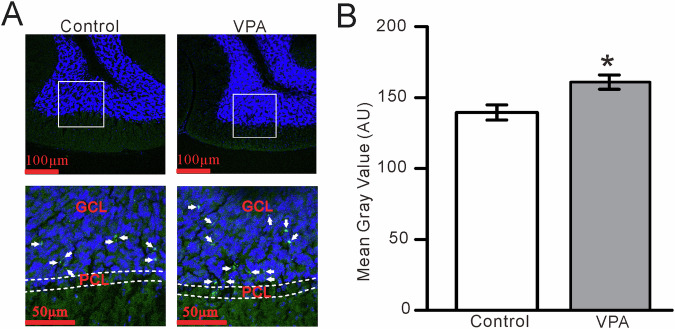
Fig. 7. GluN2A subunit-containing NMDA receptor expression in the cerebellar GC layer is increased in VPA-exposed mice compared with control mice.
A (upper panel) Digital micrographs showing confocal images of the cerebellar Crus II lobule of control (left) and VPA-exposed (right) mice. The nuclei are stained with DAPI (blue). (Lower panel) Higher magnifications of the boxed areas in (Upper panel) showing GluN2A subunit-containing NMDA receptor immunoreactivity in the GL (Green; arrows). PCL Purkinje cell layer, GCL granule cell layer. B Mean gray values of GluN2A subunit immunoreactivity in the cerebellar Crus II lobule in control and VPA-exposed mice. *P < 0.05, VPA versus control.

