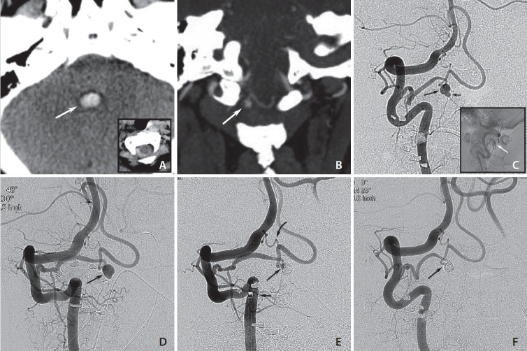
Fig. 1.
(A) Axial computed tomography (CT) scans on admission show blood in the fourth ventricle (white arrow) and subarachnoid hemorrhage at the craniocervical junction (inset). (B) Coronal CT angiography shows a small saccular structure at the C1-level (white arrow) that appears to arise from a posterior inferior cerebellar artery (PICA) with an extradural origin. (C) Right vertebral artery injections show a PICA arising from the V3-segment with an intradural aneurysm (long arrow) carrying a small bleb (short arrow). Lateral non-subtracted image shows the location of the aneurysm in the spinal canal (white arrow; inset). (D) Follow-up 9 months after aneurysm rupture shows a subtle change in shape with the bleb less clearly visible (arrow). (E) Control run after aneurysm coiling (3 coils) with a small neck remnant (long arrow), intentionally left to preserve PICA flow. Position of distal access catheter that facilitated distal microcatheter navigation into the PICA and the aneurysm (short arrow). Small anastomotic branch arising from the usual PICA origin of the V4 segment (curved arrow). (F) Follow-up angiogram 3 months after treatment shows progression of aneurysm occlusion that is now complete (arrow), while flow in the distal PICA is preserved.

