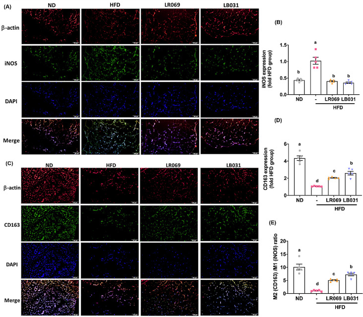
Figure 5.
LR069 and LB031 regulate M1/M2 adipose tissue macrophages (ATMs) in perigonadal adipose tissue. (A) Immunofluorescence staining of β-actin (red), M1 marker iNOS (green), and nuclei (DAPI, blue) (200× magnification; length of the scale on the right = 100 μm). (C) Immunofluorescence staining of nuclei (blue), β-actin (red), and M2 marker CD163 (green) (200× magnification; length of the scale on the right = 100 μm). (B, D) Quantification of iNOS and CD163. (E) M2 (CD163)/M1 (iNOS) ratio. The immunofluorescence staining was quantified using ImageJ software. Data are expressed as the means ± SEM. Values with different letters (a−d) differ significantly (p < 0.05) among the compared groups.

