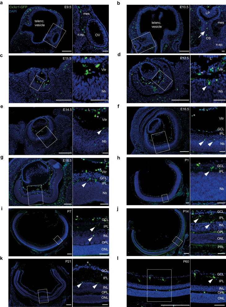
Fig. 2.
Histological analysis of the spatiotemporal development of myeloid cell populations in the murine eye. a–g Cryosections from Cx3cr1-GFP mice at different time points during prenatal development. GFP+ myeloid cells can be found in the periocular mesenchyme as early as embryonic day (E) E9.5 and enter the optic cup through the optic stalk at E10.5 (arrow), whereas hyalocytes (asterisks) can be distinguished from microglia (arrowheads) for the first time at E11.5 by localization. Scale bar = 200 µm (overview) or 50 µm (magnification). h–l Representative images from cryosections of Cx3cr1-GFP mice during early postnatal development. Hyalocytes can be found in the vitreous (asterisk), retinal microglia (arrowheads) are evenly distributed across the emerging plexiform layers. Scale bar = 200 µm (overview) or 50 µm (magnification). telenc. vesicle telencephalic vesicle, mes—mesenchyme, n.ep.—neuroepithelium, OV—optic vesicle, OS—optic stalk, LP—lens placode, L—lens, Nb—neuroblast layer, Vitr—vitreous, GCL—ganglion cell layer, IPL—inner plexiform layer, INL—inner nuclear layer, OPL—outer plexiform layer, ONL—outer nuclear layer, PRL—photoreceptor layer. Images are representative for n = 2 mice per time point

