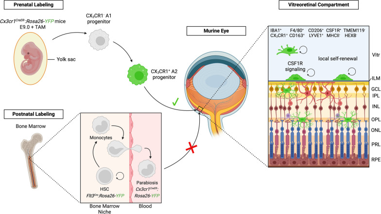
Fig. 7.
Origin, turnover and phenotype of murine hyalocytes. Graphical summary of the findings in this study. Prenatally, the local hyalocyte and microglial pool is recruited from yolk sac-derived CX3CR1+ A2 progenitors that are labeled with YFP (green) in Cx3cr1CreER:Rosa26-YFP mice after tamoxifen (TAM) injection at E9.0 and enter the vitreous cavity through the blood stream. Postnatally, fate mapping in adult Cx3cr1CreER:Rosa26-YFP mice has shown that hyalocytes, which exhibit a unique immunophenotype and rely on CSF1R-signaling for their maintenance, are long-living cells independent of replenishment from circulating peripheral myeloid cells from the definitive hematopoiesis. Hyalocytes are largely maintained by local self-renewal and reside above the inner limiting membrane while retinal microglia are located below in the neuroretina. The inner limiting membrane is constituted of both vitreal and retinal laminae. The vitreal side has a dense collagen fibril meshwork connected via extracellular matrices to the retinal glia limitans, built by the endfeet of the astrocytes (purple) residing in the nerve fiber and ganglion cell layer, separating hyalocytes and retinal microglia in two distinct compartments of the eye. Vitr—vitreous, ILM—inner limiting membrane, GCL—ganglion cell layer, IPL—inner plexiform layer, INL—inner nuclear layer, OPL—outer plexiform layer, ONL—outer nuclear layer, PRL—photoreceptor layer, RPE—retinal pigment epithelium

