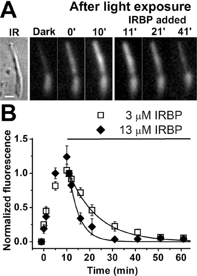
Figure 5.

Removal of all-trans retinol from human rod photoreceptor outer segments by IRBP. (A) Removal of all-trans retinol formed after rhodopsin bleaching by 3-µM IRBP. IR, infrared image of a single human rod photoreceptor (donor age 69 years). Scale bar: 5 µm. Bleaching was carried out between t = −1 and 0 minute, and IRBP was added 10 minutes after bleaching. Fluorescence images of the cell (excitation, 360 nm; emission, >420 nm) are shown at the same intensity scaling. (B) Removal of all-trans retinol by 3-µM IRBP (⬜; n = 8; donor age 69 years) and 13-µM IRBP (◆; n = 5; donor age 82 years) added at t = 10 minutes after the bleaching of rhodopsin. Retinol outer segment fluorescence intensities have been normalized over the value at t = 11 minutes, 1 minutes after the addition of IRBP. Error bars denote standard errors. The lines are simple exponentials, e–k·(t–11), decaying to 0 with unitary amplitude at t = 11 minutes, with rate constants k determined by the data points. The rate constants were 0.08 ± 0.01 min–1 for 3-µM IRBP and 0.22 ± 0.04 min–1 for 13-µM IRBP. All experiments were conducted at 37°C.
