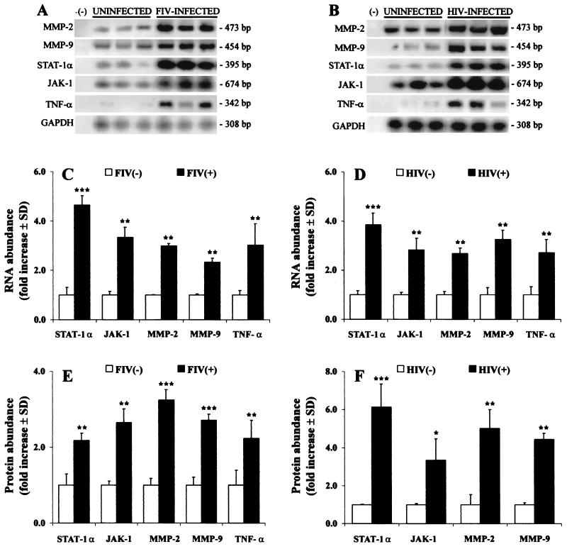
FIG. 6.
Representative MMP, TNF-α, and STAT/JAK mRNA and protein levels in brain tissue from HIV-infected persons (A, B) or FIV-infected felines (C, D) with uninfected controls. RT-PCR showed increased MMP-2 and -9, STAT-1, JAK-1, and TNF-α mRNA levels in FIV-infected feline (n = 4) (A, C) and HIV-infected human (n = 5) (B, D) brain tissue compared to brain tissue from uninfected felines (n = 4) and human controls (n = 6). Amplification of GAPDH was used to ensure equal template loading. Gelatin zymography and Western blot analysis revealed concurrent increases in STAT-1α, JAK-1, MMP-2, and MMP-9 in FIV-infected (E) and HIV-infected (F) brains compared to uninfected controls. In contrast to macrophages, only the STAT-1α monomer was detected in brain tissue. Data are expressed as fold increases over uninfected controls and represent the mean ± standard deviation of three experiments. Significant differences relative to uninfected controls are indicated (Tukey-Kramer test; ∗, P < 0.05; ∗∗, P < 0.01; ∗∗∗, P < 0.001).

