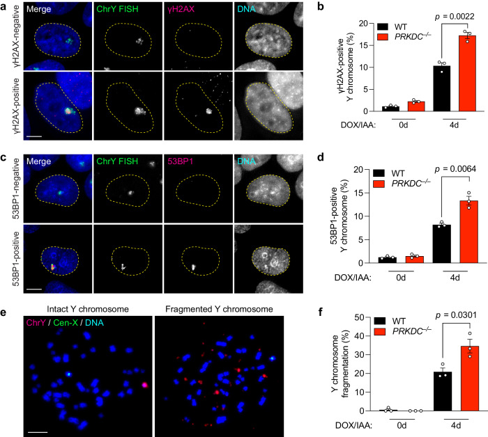
Fig. 3. NHEJ-deficient cells accumulate damaged chromosome fragments within MN bodies in the nucleus.
a Images of interphase cells with γH2AX-negative Y chromosome or γH2AX-positive Y chromosomes within an MN body after 4d DOX/IAA treatment. Scale bar, 5 µm. b Frequency of Y chromosomes marked by extensive γH2AX in the nucleus. Data were pooled from (left to right): 549, 353, 429, and 293 cells. c Images of interphase cells with 53BP1-negative or 53BP1-positive Y chromosomes after 4-day DOX/IAA treatment. Scale bar, 5 µm. d Frequency of Y chromosomes marked by extensive 53BP1 in the nucleus. Data were pooled from (left to right): 567, 543, 634, and 592 cells. e Images of metaphase spreads with an intact or fragmented Y chromosome after 4d DOX/IAA treatment. Scale bar, 10 µm. f Frequency of Y chromosome fragmentation. Data pooled from (left to right): 329, 291, 347, and 373 metaphase spreads. Bar graphs in (b), (d), and (f) represent the mean ± SEM of n = 3 independent experiments. Statistical analyses were calculated by two-tailed unpaired Student’s t-test. Source data are provided as a Source Data file.

