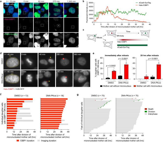
Fig. 4. Inhibition of NHEJ prolongs 53BP1 residence time at MN bodies and triggers cell cycle arrest.
a Example time-lapse images of a mother cell harboring a dCas9-SunTag-labeled Y chromosome in a micronucleus undergoing cell division, which in turn generates a daughter cell that incorporates the micronucleated chromosome into the nucleus as an MN body labeled with a HaloTag fused to the minimal focus-forming region of 53BP1 (Halo-53BP1). Time point at 0 min depicts mitotic entry. Scale bar, 5 µm. b Measurement of Halo-53BP1 fluorescence intensity over a ~37-h imaging period. The dCas9-SunTag-labeled Y chromosome is monitored as a control. c Schematic of Halo-53BP1 residence time at MN bodies from (a, b) after mitosis. d Representative time-lapse images of newly-formed, Halo-53BP1-labeled MN bodies with or without treatment with the DNA-PKcs inhibitor AZD7648. Scale bar, 5 µm. e Frequency of Halo-53BP1-labeled MN body formation and persistence. Data represent mean ± SEM of n = 3 independent experiments pooled from (left to right): 63, 64, 72, and 74 micronucleated mother cells. Statistical analyses were calculated by ordinary one-way ANOVA test with multiple comparisons. f Duration of Halo-53BP1 residence time from (d) after MN body formation with or without inhibition of DNA-PKcs in individual daughter cells. g Fate of daughter cells after division of micronucleated mother cells. Sample sizes in (f, g) represent individual daughter cells. Source data are provided as a Source Data file.

