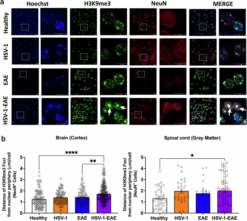
Fig. 7. H3K9me3-associated SAHF formation in brain and spinal cord neurons of mice with asymptomatic HSV-1 brain infection and EAE.
Brain and spinal cord tissues from four animals per group were harvested 14 days after EAE induction, 45–50 days after HSV-1 infection, or mock-treatment alone for detecting the expression of H3K9me3 foci by immunofluorescence. a Representative images showing Hoechst nuclei staining (blue), H3K9me3 staining (green), NeuN marker (red), and image merges. Left: images in each fluorescence channel correspond to 100X magnifications, and right: Images are shown at a 5X optic zoom of the area outlined in squares with white dashed lines. Scale bars = 10 μm. White arrows show senescence-associated heterochromatin foci (SAHF). b Quantification of the distance of each H3K9me3 foci from the nuclear periphery in NeuN+ cells. Values represent means ± SEM of the measurements carried out in at least ten fields in the brain tissues and three fields in the spinal cord tissues per sample. Data were analyzed using One-way ANOVA followed by Bonferroni’s post-hoc test for analysis from brain tissues and Kruskal–Wallis followed by Dunn’s post-hoc test for analysis from spinal cord tissues; ****p < 0.0001, **p < 0.01, *p < 0.05. White bars with circles represent data from healthy mice, orange bars with triangles represent data from mice with HSV-1 infection, blue bars with diamonds represent data from mice with EAE and purple bars with squares represent data from mice with EAE and HSV-1 infection.

