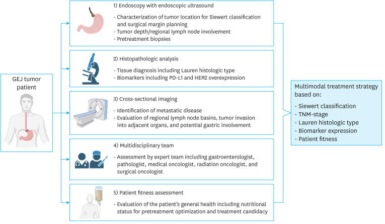
Fig. 3. Flowchart depicting the elements required for a comprehensive GEJA work-up including: 1) EGD/EUS for anatomic localization (Siewert classification and extent of proximal esophageal and distal gastric involvement), evaluation of tumor depth and regional LNs for TNM staging, and pretreatment biopsies; 2) evaluation of preoperative biopsies for Lauren histologic type, PD-L1 (CPS), MSI, and HER2 overexpression status; 3) CT scan ± PET scan of the chest, abdomen, and pelvis for evaluation of regional LN basins and metastatic disease for TMN staging, tumor invasion into adjacent organs, and gastric wall thickening indicating possible involvement; 4) evaluation by a multidisciplinary team; and 5) assessment of patient fitness and nutritional status for pretreatment optimization and determination of treatment candidacy.
GEJA = gastroesophageal junction adenocarcinoma; EGD = esophagogastroduodenoscopy; EUS = endoscopic ultrasonography; LN = lymph node; TNM = tumor, node, metastasis; PD-L1 = programmed cell death ligand 1; CPS = combined positive score; MSI = microsatellite instable; HER2 = human epidermal growth factor receptor 2; CT = computed tomography; PET = positron emission tomography; GEJ = gastroesophageal junction.

