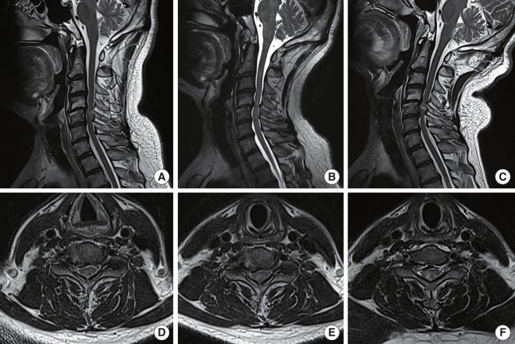
Fig. 4.

Increase of SI grade and cervical stenosis during extension. A 53-year-old male was diagnosed with cervical spondylotic myelopathy due to a herniated cervical disc at C4–5–6. (A) Flexion-positioned magnetic resonance imaging (MRI) showed intramedullary faint SI (G1) at C4–5 and no effacement of subarachnoid space. (B) Neutral-positioned MRI showed complete obliteration of the anterior subarachnoid space (Muhle grade 2). (C) Extension-positioned MRI showed severe cord impingement at C4–5–6 (Muhle grade 3) and intense intramedullary SI (G2). (D) Flexion-positioned axial view showed cord compression, but the subarachnoid space was not obliterated. (E) On the neutral axial T2-weighted imaging (T2WI), effacement on the right anterior subarachnoid space was noted. (F) On extension-positioned axial T2WI, intense intramedullary SI (G2) was observed.
