
Figure 3.
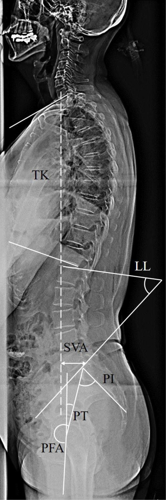
Measurement of spinopelvic parameters in the whole-spine lateral radiograph. The following parameters were measured: SVA (the distance between the plumb line passing through the center of the C7 vertebra and posterior superior corner of the sacral endplate), TK (the angle between the upper endplate of T1 and lower endplate of T12), LL (the angle between the upper endplate of L1 and the endplate of the sacrum), and PI (the angle between a line perpendicular to the endplate of the sacrum and a line drawn from the midpoint of the bilateral femoral head centers to the midpoint of the sacral endplate), PT (the angle between the plumb line and a line drawn from the midpoint of the bilateral femoral head centers to the midpoint of the sacral endplate), PFA (the angle between a line drawn from the midpoint of the bilateral femoral head centers to the midpoint of the sacral endplate, and the proximal femoral shaft axis). TK, thoracic kyphosis; LL, lumbar lordosis; SVA, sagittal vertical axis; PI, pelvic incidence; PT, pelvic tilt; PFA, pelvic femoral angle.