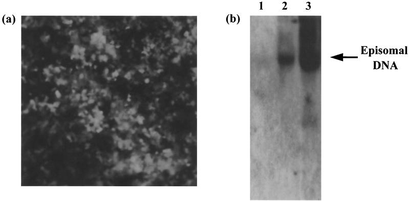
FIG. 1.
(a) Production of HVS stably transduced A549 cell line. A549 cells were infected with HVS-GFP at a MOI of 1 and cultured in the presence of Geneticin. After 2 weeks, only cells which had been successfully transduced remained viable, with 100% exhibiting the GFP phenotype when analyzed by fluorescence microscopy. (b) Gardella gel and Southern blot analysis of A549 cells (lane 1), HVS stably transduced A549 cells (lane 2), and HVS-transformed B133 T cells (lane 3). Episomal and linear DNAs were separated by electrophoresis, transferred to nitrocellulose, and hybridized with a radiolabeled 32P-labeled random-primed probe specific for the KpnE fragment of the HVS genome.

