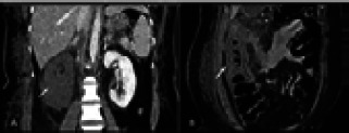
Figure 5.

36 years non diabetic female presented with severe abdominal pain with raised TLC, serum creatinine and D-dimer. (A and B) Coronal CECT sections show complete non- enhancement of right kidney (solid white arrow) with perinephric fat stranding and long-segment thickening and hypo-enhancement of ascending colon (solid white arrow) with peri-colonic fat stranding suggesting ischemic changes. Histopathology done on operative specimen showed angio-invasive fungal profiles suggesting mucormycosis
