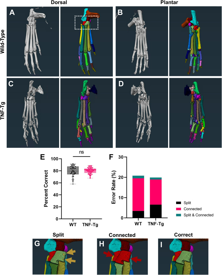
Fig 1. Semi-automated segmentation of TNF-Tg hindpaws with severe arthritis shows comparable accuracy to wild-type counterparts.
To evaluate bone-specific erosions in the TNF-Tg mouse model of inflammatory-erosive arthritis, we utilized our recently published high-throughput semi-automated hindpaw segmentation protocol [46]. Representative images of the dorsal and plantar surfaces of hindpaw micro-CT images (left) and the segmentation of each bone indicated by unique colors (right) are provided for wild-type (A-B) and TNF-Tg (C-D) male mice at 8-months of age. Without user intervention, the semi-automated protocol produces accurate segmentations of approximately 80% of bones in wild-type datasets (error rate ~20%), which remarkably remains consistent, and potentially improved through reduced variance, for TNF-Tg mice with bone erosions (E). Note that TNF-Tg segmentations have increased split and reduced connected errors, corresponding with eroded bones (F). If present, these errors are corrected utilizing described workflows [46] to produce the final correct segmentation. Examples of errors and correction (G-I) are provided from a high-magnification image of the tarsal region in the wild-type hindpaw shown in A (white box).

